Medical Diagnostics At The Horder Centre

Medical Diagnostics At The Horder Centre

Accurate diagnosis is essential to getting the right treatment for your health problem.

That’s why, at The Horder Centre, our experts use the best tools available to ensure your conditions are diagnosed correctly.

We have a range of different diagnostic services available at The Horder Centre for insured, NHS and self-pay patients.

Types of diagnostic services at The Horder Centre

The three main tools used for our diagnostic services at The Horder Centre are magnetic resonance imaging (MRI) scans, ultrasounds and X-rays.

These tried and tested tools are the foundation of accurate diagnostic testing.
MRI

Magnetic resonance imaging (MRI)

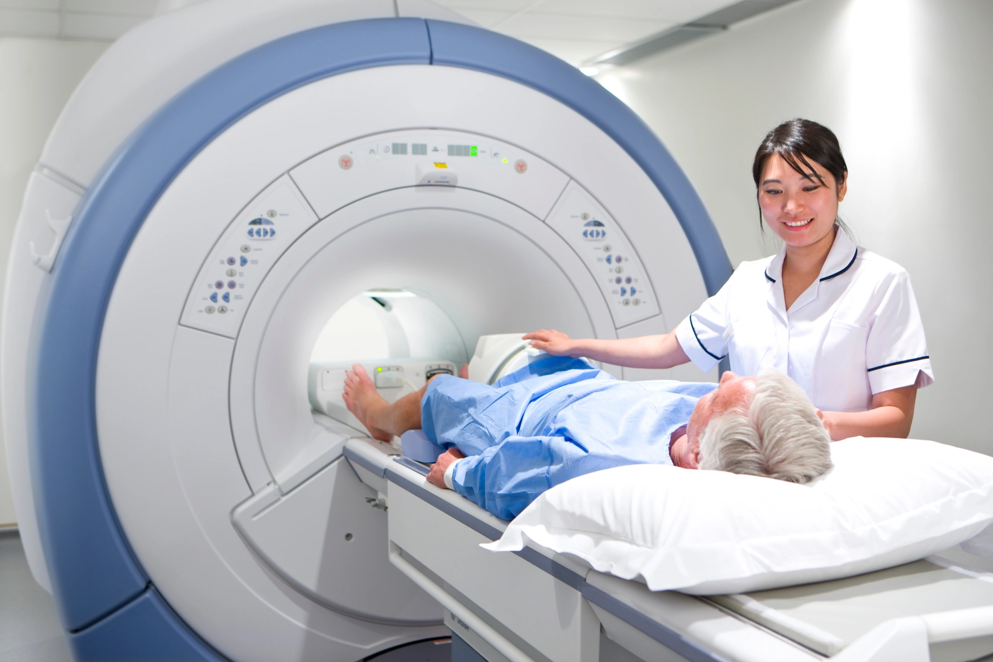
During magnetic resonance imaging (MRI), a large machine uses a magnetic field and radio waves alongside a computer to create a series of images that show a specific area of the body that needs to be investigated. These detailed images can be used to see your bones and soft tissues, helping us to make an accurate diagnosis.

Because of the level of detail they’re able to provide, MRI scans can offer a clearer picture of certain diseases and irregularities that may not be detectable on an X-ray or ultrasound.

What happens during an MRI?

Before you have an MRI, you might be given a contrast medium (dye) through an injection in your vein. This is to help the images be as clear as possible, however, it isn’t always necessary.

The scan itself will be done while you are lying down and you will either move into the scanner head or feet first, depending on what part of your body is being scanned.

The scanner is controlled by a radiographer who will sit at a computer in a separate room. They will still be able to see you through the viewing window and you can communicate with them through the intercom.

The scanner will make loud, tapping noises at specific points. This is caused by the electric current in the scanner coils being switched off and on. Because of the noise, you will be given earplugs.

You need to keep as still as possible during your scan and you might be instructed to hold your breath for a few seconds. The scan can last from 15 to 90 minutes depending on where your body is being scanned and how many images need to be taken.

ultrasound

Ultrasound

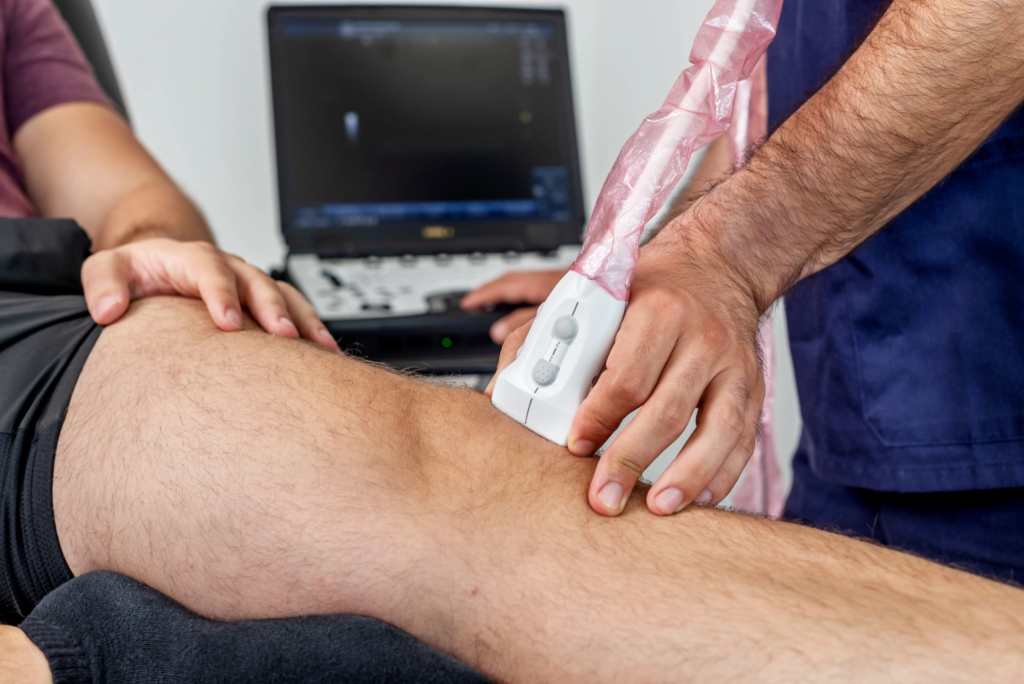
Ultrasound scans use high-frequency waves to create images of muscles, tendons, ligaments and joints.

Ultrasounds use sound waves as opposed to radiation, so they are thought to be very safe. An ultrasound probe emits these high-frequency sound waves and is pressed against your body.

These sound waves will bounce off your body and create ‘echoes’. These echoes are picked back up by the probe and are then translated into an image on a computer screen to provide us with a real-time view of an area of your body.

What happens during an ultrasound?

Before an ultrasound, you might be asked to drink lots of water to fill your bladder. This can provide a clearer view of your womb or pelvic area. Sometimes you might be asked to avoid food or drink for several hours beforehand if your digestive system needs to be viewed.

The ultrasound will be performed by one of our expert consultant radiologists.

You will be asked to lie down and a cold gel will be applied to your body. This gel helps the probe glide over your body and keeps the contact continuous for accurate imaging.

The procedure usually takes between 15 minutes and half an hour.

What can an ultrasound be used to diagnose?

  • Sprains
  • Strains
  • Tears
  • Other soft tissue conditions
xray

X-ray

An X-ray is a diagnostic technique that captures abnormalities in your bones and specific bodily tissues using radiation. The levels of radiation are low, so the risk to your health is minuscule.

X-rays are a type of radiation that passes through your body. As they do this, they are absorbed into your body at different rates. This is then picked on the other side of the body by a detector and the results are turned into images.

What happens during an X-ray?

During an X-ray, you will be asked to lie on a table or stand against a flat surface, depending on which part of your body is being assessed. The X-ray machine will be positioned and aimed at your body by the radiographer who will either be behind a screen or in the next room.

X-rays last for less than a second and you won’t feel anything while it is being done. You will need to keep still to ensure the images are not blurred.

What can an X-ray be used to diagnose?

X-rays are commonly used to diagnose:

  • Bone fractures or breaks
  • Certain tumours and other abnormal masses
  • Calcifications (a build-up of calcium in your tissue, blood vessels or organs which causes them to harden)

Contact The Horder Centre

At The Horder Centre, our highly skilled team use all three of these accurate diagnostic tools, depending on what we’re looking for, to make sure you receive the best treatment for your condition.

If you’re concerned about your health and want to use our diagnostic services, contact us today. We’ll help you get to the bottom of your health problems and let you know which treatment is right for you.

Orthopaedic treatment at The Horder Centre

We offer a range of high quality, specialist services led by the very best clinicians and health professionals in their relevant fields.

What makes Horder Healthcare unique

Horder Healthcare is committed to providing the very best quality of care for our patients and customers. We are continuously working on improving and reducing risks and this is reflected in our consistently high CQC results, patient satisfaction questionnaires and minimal levels of infection.

Latest news and articles

hip-replacement

Consultant Q&A: Mr Senthil Velayudham

Image of nutritious food

Nutrition and Hydration Week 2024

Banner women health check

The McIndoe Centre introduces Women’s Health Check 

We are a charity

We reinvest our profit to benefit more people and help us achieve our aim of advancing health.

fm_weareacharity2